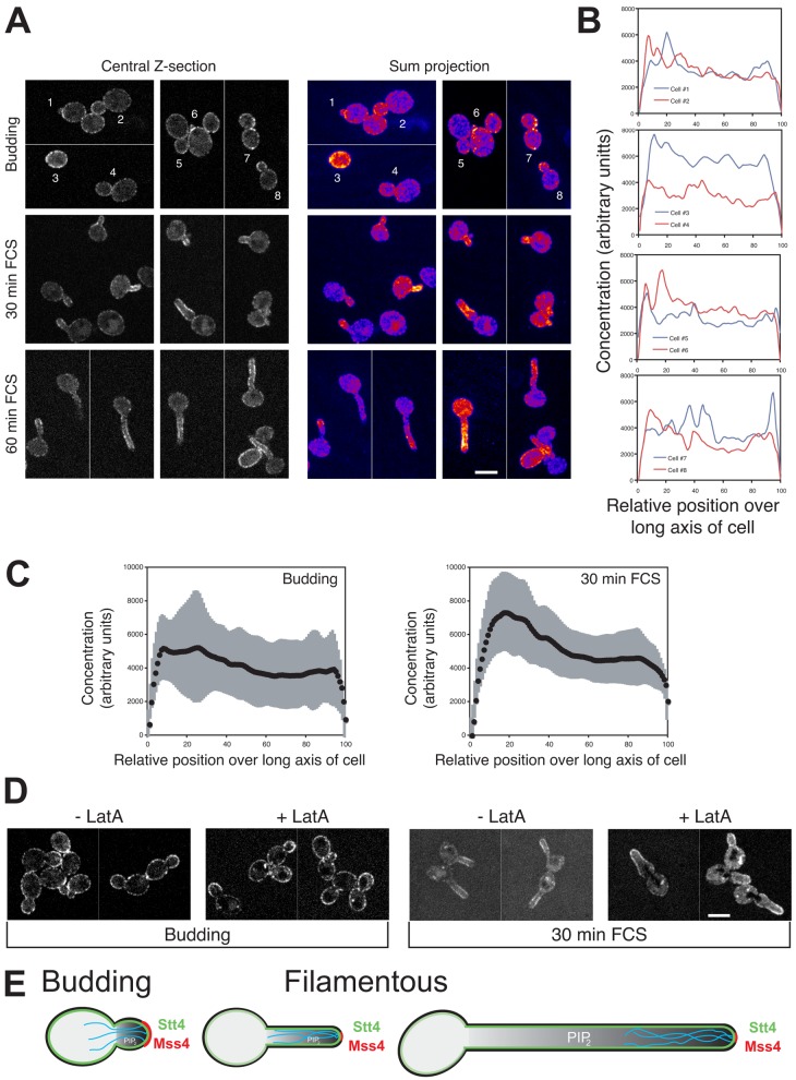
Figure 10.
Stt4 localizes to punctae on the plasma membrane, independent of the actin cytoskeleton. (A) Stt4 is localized to the plasma membrane. A stt4 strain expressing GFP-Stt4 was grown in the presence of Dox. Central z-sections and false-colored sum projections (21 z-sections) of representative cells grown as indicated are shown (ni.ex. = 3). (B) Quantification of Stt4 concentration over long axis of budding cells. Signal concentration over the long axis of budding cells from A, as described in Fig. 4 A. (C) Average GFP-Stt4 signal concentration over the long axis of budding cells or cells incubated with FCS as described in Fig. 4 A (n = 41 cells from A) is shown with SD in gray. (D) F-actin is not required for plasma membrane distribution of GFP-Stt4. Strains as in A were treated with LatA, described in Fig. 6 A. Central z-sections of representative cells are shown (ni.ex. = 2). (E) Schematic representation of the mechanisms involved in generating and maintaining long-range PI(4,5)P2 gradient in filamentous cells. PI(4,5)P2 gradient shown (gray) in cell with actin cables (cyan) and Mss4 (red) and Stt4 (green) indicated.

