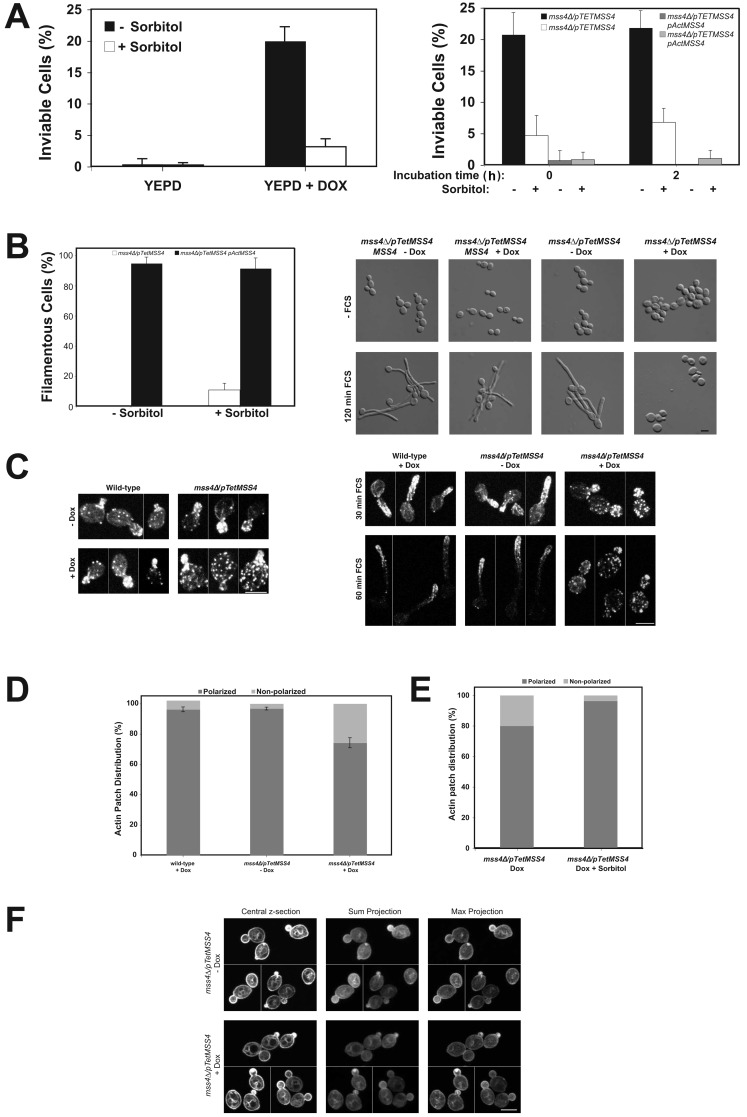
Figure 2.
The mss4 filamentous growth defect is not due to perturbation of the actin cytoskeleton or altered Cdc42 localization. (A) Inviability of mss4 mutant is suppressed by sorbitol and unaffected by FCS. The mss4 strain (left) or indicated strains (right) were grown in the presence or absence of Dox, with and without 0.5 M sorbitol, and incubated with FCS for indicated times (right). Average percentage of inviable cells (ni.ex. = 3; n = 4 determinations; 75 cells each [left]; and ni.ex. = 2; 10 times 50 cells each [right]). (B) Inviability of mss4 mutant is not responsible for filamentous growth defect. The percentage of filamentous cells after 2 h in FCS from A (left). Images of indicated cells grown with 0.5 M sorbitol (ni.ex. = 3; right). (C) The actin cytoskeleton is disorganized in mss4 cells. Actin cytoskeleton in indicated strains grown with or without Dox incubated with FCS. Maximum projections (4–8 z-sections [left]; 6–9 z-sections [right]) of representative budding cells from different fields of view (left; ni.ex. = 2). (D) Quantitation of actin patch distribution in budding mss4 cells. Averages indicated with bars showing values (ni.ex. = 2; 150 cells each). (E) Sorbitol restores polarized actin patch distribution in mss4 cells. Actin patch polarity was determined in indicated budding cells (ni.ex. = 2; n = 160 cells in the absence and presence of 0.5 M sorbitol). (F) Cdc42 localization is unaffected in the mss4 mutant. Central, sum, and maximum projections (10 z-sections) of representative mss4 cells expressing GFP-Cdc42 from different fields of view. A cluster of GFP-Cdc42 was apparent within the bud of some cells (+ Dox; ni.ex. = 2).

