Figure 1.
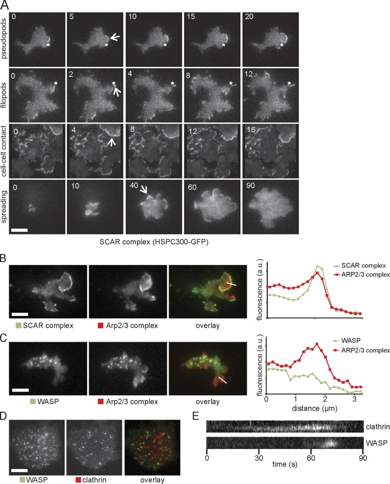
SCAR complex and WASP localization in wild-type D. discoideum cells. (A) TIRF microscopy of SCAR complex (labeled with HSPC300-GFP) in pseudopods, filopods, cell–cell contact, and cell spreading of wild-type cells. Numbers indicate the time in seconds. The position of the asterisks is fixed across different time points. Arrows show SCAR-rich protrusions. (B and C) Colocalization of GFP-tagged SCAR complex (B) and WASP (C) with RFP-tagged Arp2/3 complex (subunit ARPC4) during cell migration. Quantitations of the fluorescence intensity along the indicated lines through the pseudopod are displayed on the right. Images are representative of ≥50 cells observed. (D) Colocalization of GFP-WASP and RFP-clathrin light chain. (E) Kymograph from a video similar to D showing arrival and disappearance of clathrin and WASP during a single clathrin-mediated endocytosis event. a.u., arbitrary unit. Bars, 5 µm.

