Figure 2.
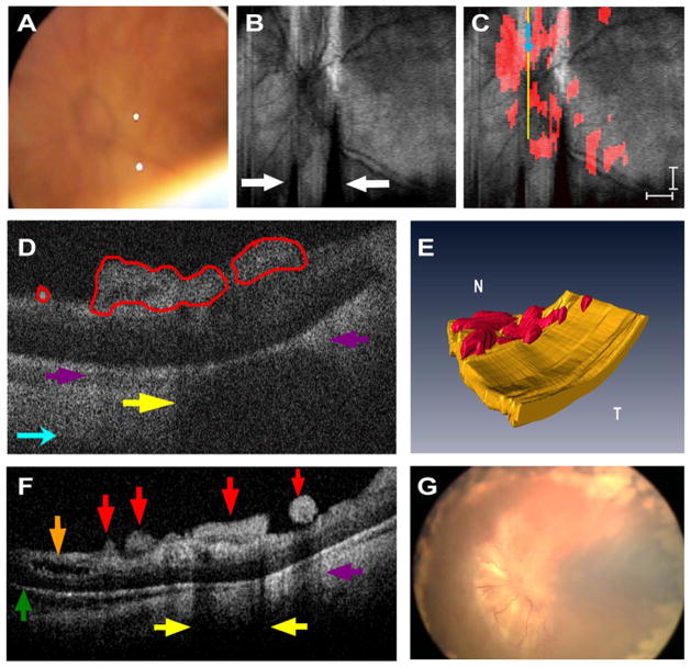
Patient 1’s left eye. A, Video-indirect image of the optic nerve and posterior pole of the retina without evidence of preretinal structures. B, Sum voxel projection image of optic nerve and posterior pole. There is also partial loss of some B-scan images from motion artifact (white arrows). C, Same summed voxel projection image in (B) with preretinal tissue (red areas in D) surrounding and overlying optic nerve. Light blue arrows provide image orientation (B, C). Scale bars are 1000 μm. D, Cropped cross-sectional spectral domain optical coherence tomography (SD OCT) image demonstrates preretinal structures traced in red and corresponds to yellow vertical line in (C). Purple arrows denote shadowing artifact of preretinal structures (D, F), and yellow arrows point to focal shadowing (D, F). E, Three-dimensional SD OCT image depicts preretinal structures in the posterior pole. N = nasal; T = temporal. F, Representative cross-sectional image demonstrates a variety of subclinical pathology. Green arrow indicates retinal detachment, brown arrow denotes retinoschisis, and red arrows point to preretinal structures. G, Retcam fundus photograph demonstrates tractional retinal detachment 1 month after SD OCT imaging.

