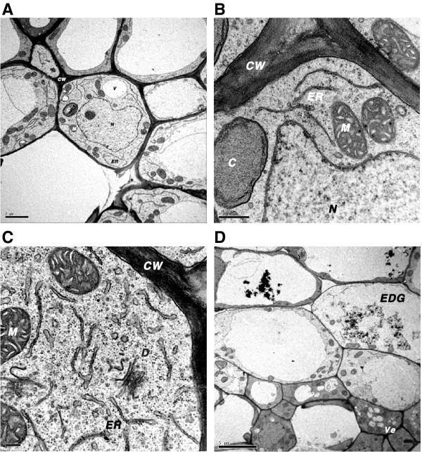
Figure 4.
Transmission electron micrographs of rice cells expressing bp100der transgenes.(A) Control Senia vascular and surrounding, and parenchyma cells of the crown region showing normal morphology. (B) Detail of endoplasmic reticulum morphology of Senia cells. (C) Cells surrounding vascular cells, showing increased abundance of dictysome vesicles and distinct dilation of ER cisterna in S-bp100.2i cells. (D) Vesicles in S-bp100.2mi cells surrounding vascular cells and accumulation of electron dense granules in parenchyma cells. CW, cell wall; D, dictysome; EDG, electron-dense granules; ER, endoplasmic reticulum; M, mitochondria; N, nucleus; Ve, vesicle. Scale bars: 2 μm (A), 0.5 μm (B), 0.2 μm (C) and 5 μm (D).

