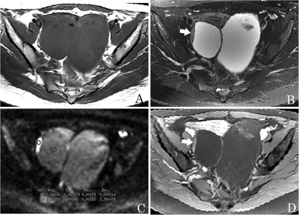
Figure 3.

A 28-year-young woman with bilateral benign serous ovarian cystadenomas. (A) An axial T1-weighted image shows a pelvic tumor with hypointensity. (B) Axial T2-weighted image shows a complex adnexal tumor with solid and cystic components, and solid components as vegetations with intermediate signal intensity (arrow). (C) Axial diffusion-weighted imaging (DWI) obtained at b =1,000 s/mm2 shows the solid component as hyperintense with a high apparent diffusion coefficient (ADC) value (circle 1, ADC = 1.26 × 10⁻³ mm2/s), and the cystic component with an even higher value (circle 2, ADC = 2.59 × 10- 3 mm2/s). (D) Axial contrast-enhanced T1-weighted image demonstrates slight enhancement of the solid component (arrow).
