
Figure 1.
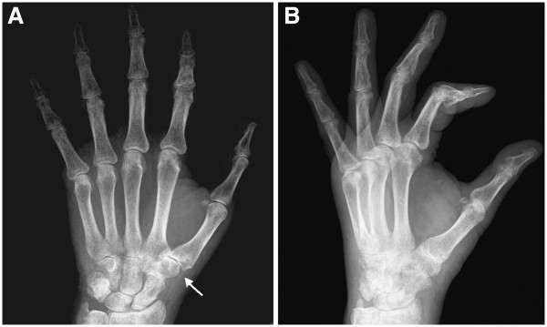
Plain radiograph of the hand performed after the second clinical examination. (A) Anteroposterior view. (B) Lateral view. Space narrowing of the trapeziometacarpal and trapezionavicular joints (arrow in [A]). Diffuse osteoporotic picture of the carpal and the phalangeal bones, to be interpreted as disuse osteoporosis.