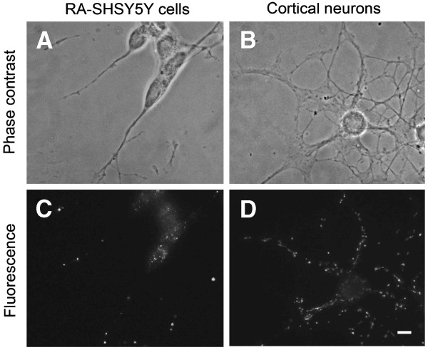
Figure 1.
Aβ42 immunoreactivity in retinoic acid differentiated neuroblastoma cells and neonatal rat cortical neurons. Phase contrast and fluorescence (greyscale) images of RA differentiated SH-SY5Y human neuroblastoma cells (A, C) and cultured rat cortical neurons (B, D), respectively. Aβ42 positive granules are bigger and more abundant in neurites than in perikarya. Bar, 50 nM.

