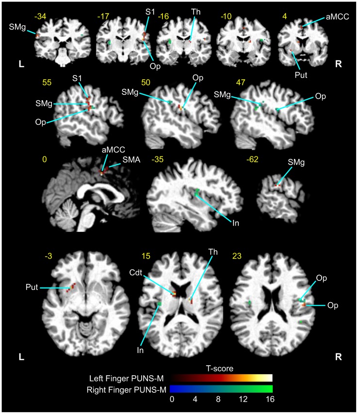
Figure 4. PUNS-M stimulates BOLD contrast signals in somatosensory detection and discrimination networks.
Grand average fMRI BOLD responses obtained from five subjects in response to left finger (red-yellow LUT) and right finger (blue-green LUT) stimulation with PUNS-M waveforms (L = Left, R = right). Anatomical areas shown were significantly activated (p<0.001). The yellow numbers correspond to MNI slices in respective views while white labels indicate anatomical regions abbreviated as follows: SMg = supramarginal gyrus; S1 = primary somatosensory cortex; Op = parietal operculum; Th = thalamus; aMCC = anterior middle cingulate cortex; Put = putamen; SMA = supplementary motor area; In = Insula; Cdt = Caudate.

