Figure 1.
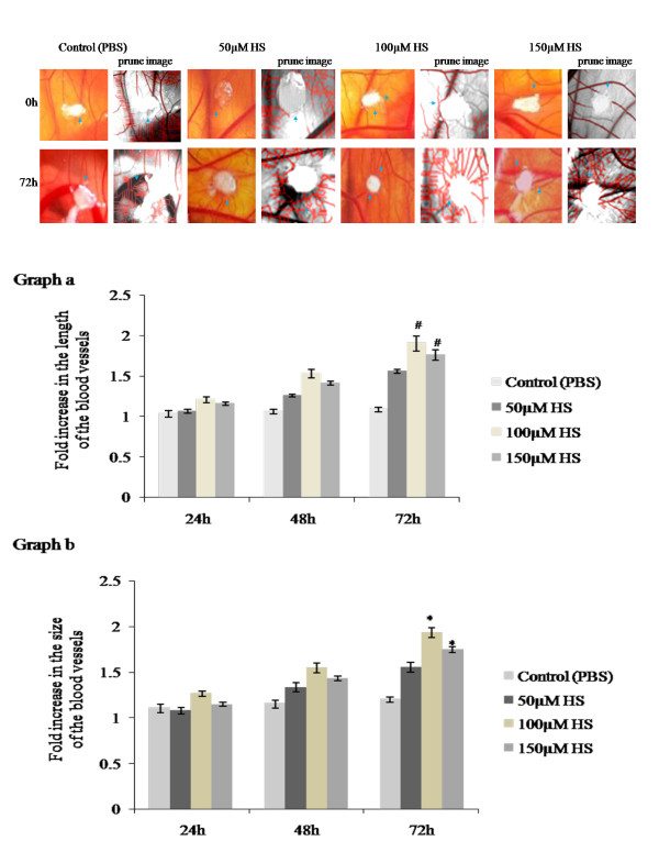
a. Effect of biotin conjugated heparin on late CAM. Images of late CAM treated with 50, 100 and 150 μM heparin. Control CAM was treated with 1X PBS. CAM treated with 100 μM heparin shows increased vascularization around the sponge as spoke wheel pattern which is comparatively higher than other concentrations and is also supported by skeletonized prune images by Angioquant software. Images are taken using Canon digital camera at 4X magnification and are the result of 3 different set of experiments. Arrow indicates the presence of blood vessels. 1b. Effect of biotin conjugated heparin on the growth of blood vessels on late CAM. Total length and size of the blood vessels was measured using Angioquant software after treating CAM with 50, 100 and 150 μM heparin. Control CAM was treated with 1X PBS. Images recorded at 0, 24, 48 and 72hours were analyzed individually. Values at 0hour were taken as one for all. CAM treated with 100 and 150 μM heparin shows significant increase in vessels length (graph a) and size (graph b) after 72hours of treatment when compared to control. Though both the concentrations shows significant changes, 100 μM heparin shows comparatively higher angiogenic potential. Experiments were performed in triplicate and data presented as mean ± SEM, #p = 0.001 and *p = <0.001 versus control.

