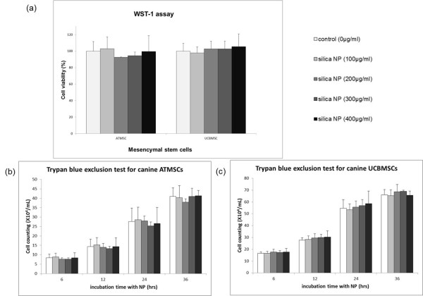
Figure 3.
Determination of cell viability and proliferation in the presence of silica nanoparticles for labeling of canine MSCs. (a) Canine MSCs were incubated without (control) or with 100, 200, 300, or 400 μg/ml silica nanoparticles (NP) for 24 h, and then cell viability was measured by a WST-1 assay. No significant differences in metabolic activity were observed compared with that of the control during culture with silica nanoparticles. Each concentration was assayed in triplicate. (b, c) Trypan blue exclusion showed that the proliferation of canine MSCs was not inhibited. No significant differences in cell number were observed compared with that of the control during culture with silica nanoparticles. Each concentration was evaluated in triplicate. Data are the means ± standard deviations (P > 0.05, analysis of variance).

