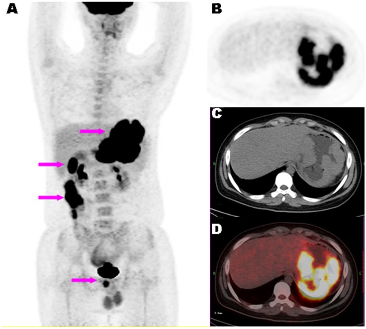
Figure 2. A representative case of ANHL.
. 18F-FDG PET/CT images of a15-year-old male patient with newly diagnosed Burkitt lymphoma. Maximum-intensity-projection view (A) showing multiple hypermetabolic lesions in the gastrointestinal tract (arrows). Axial PET (B), CT (C) and fused PET/CT (D) images showing diffused and irregular thickening of gastric wall with FDG uptake (SUVmax of 18.82).

