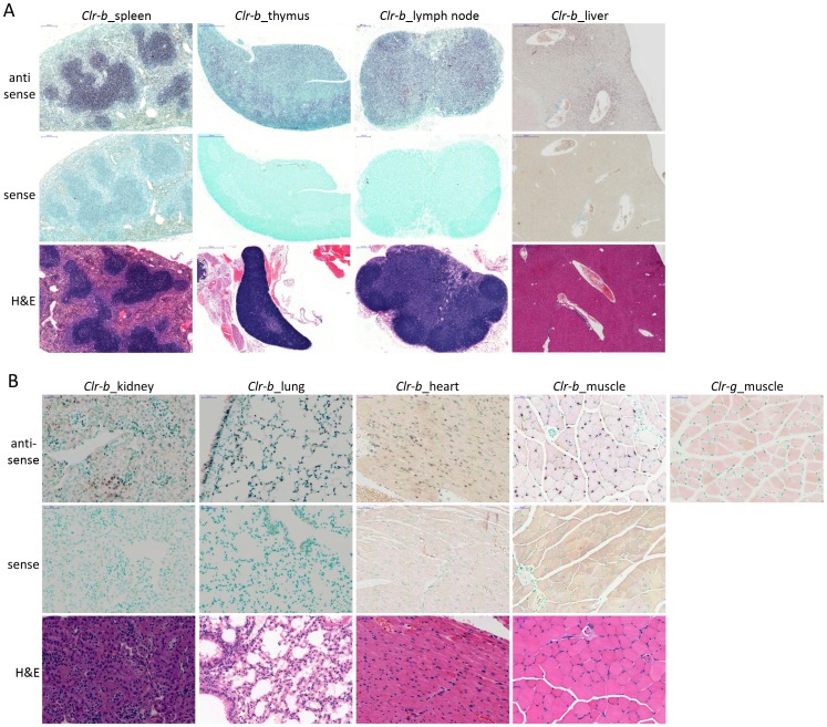
Figure 8. Wide-spread expression of Clr-b in lymphoid and non-lymphoid tissues and cell types.
In situ hybridization was performed on tissue sections from a PBS-perfused B6 mouse. Hybridization of the indicated (A) primary and secondary lymphoid organs and (B) non-lymphoid organs was performed with a DIG-labeled anti-sense Clr-b RNA probe and revealed with an alkaline phosphatase-conjugated anti-DIG secondary mAb. Control in situ hybridization with a DIG-labeled Clr-b sense RNA probe is shown. Anti-sense Clr-g staining of hind leg muscle tissue is provided as a hybridization specificity control. H&E staining is provided to identify cell types and reveal organ structure. The scale bars indicate 200 µm in the lymph node and spleen, 500 µm in the liver and thymus, and 50 µm in the kidney, lung, heart, and muscle.

