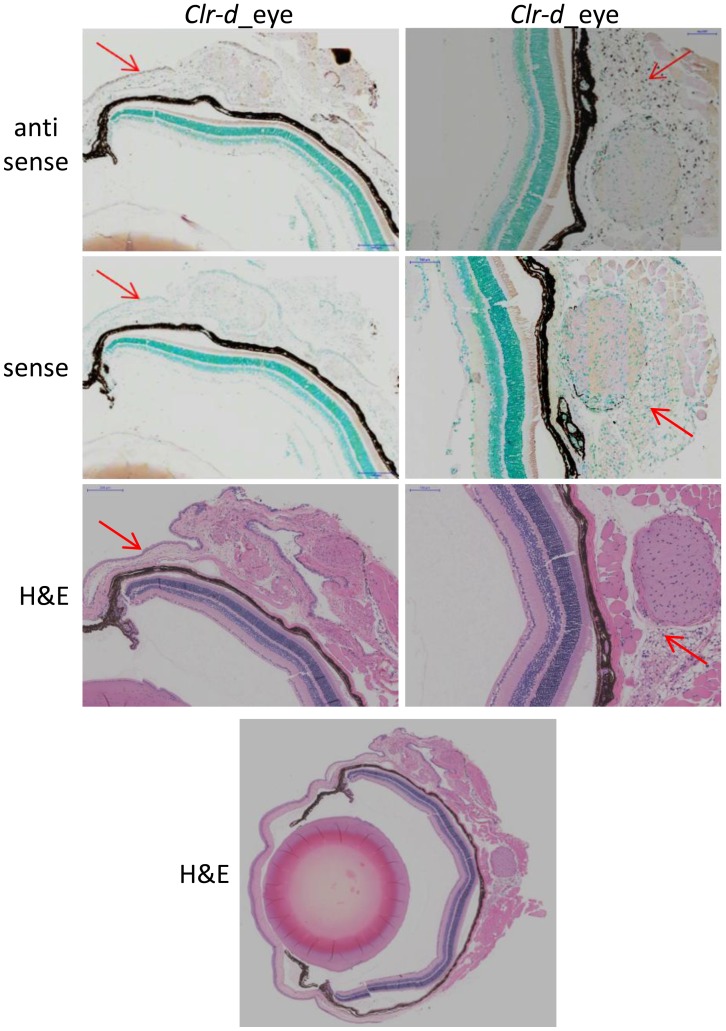
Figure 9. Clr-d is expressed in distinct areas of the mouse eye.
A DIG-labeled RNA probe for Clr-d was used to identify regions of the eye expressing this gene. Expression was detected in the corneal epithelial progenitor cells (left column) and the cell mass surrounding the optic nerve (right column). Red arrows indicate areas of positive staining. Sense (control) RNA hybridizations are shown along with H&E stains to elucidate eye cell types and architecture. For organ orientation an H&E stain of the whole eye is provided at the bottom. The scale bar in the sclera epithelial cell section indicates 200 µm, and 100 µm in the optic nerve section.

