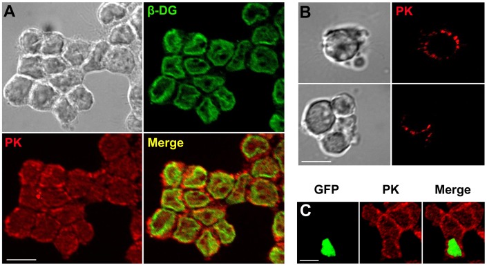
Figure 2. Localization of endogenous pikachurin to the cell surface in Y79 cells.
(A) Immunofluorescent images of pikachurin (PK, red) and β-DG (green). Most pikachurin label is present on the extracellular side of the immunolabeling for DG, predominantly a membrane bound protein. (B) Live imaging of pikachurin in cells shows punctuate extracellular staining. (C) Overexpression of dystroglycan in transfected Y79 cells, indicated by the nuclear GFP, had no significant influence on pikachurin staining. A representative field of view is shown here, and another example can be found in Fig. S1. Bar = 10 µm.

