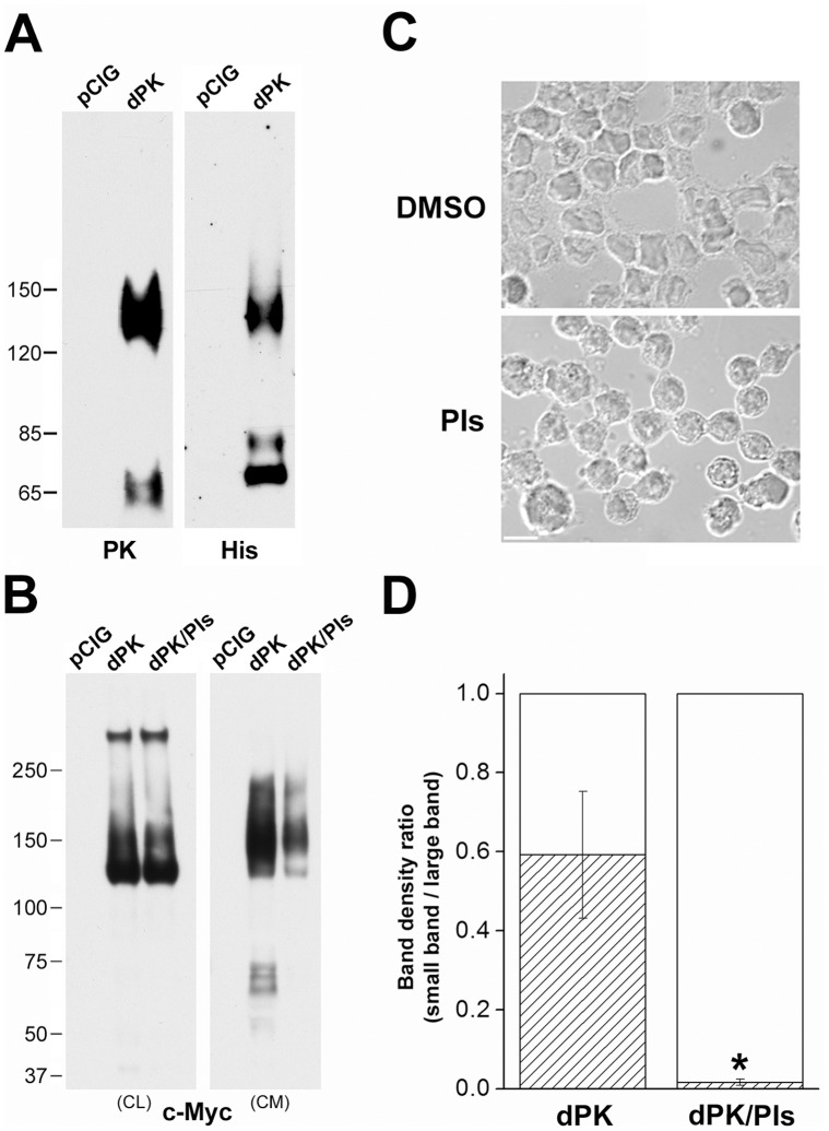
Figure 4. Pikachurin fragments present in the conditioned medium of Y79 cells.
(A) Western blots of the conditioned medium from pCIG and dPK transfected cells. PK antibody labeled N fragments and whole protein and His antibody labeled C fragments and whole protein. (B) Inclusion of protease inhibitors in culture medium (dPK/PIs) didn’t affect the expression of dPK in the cell lysates (CL) but decreased its levels in the conditioned medium (CM). Note: c-Myc labels only recombinant protein. Short exposure reveals the high levels of recombinant pikachurin monomers and oligomers on the CL blot. Small fragments on this blot were visible with prolonged exposure but on a much darker background (data not shown). (C) Morphological changes in Y79 cells induced by incubation with protease inhibitors for 48 hrs. The solvent DMSO was diluted 1∶400 in the culture medium for control cells. Bar = 10 µm. (D) Density ratios of Western blot bands (N-fragments/monomers, Fig. 4B). N = 3 experiments. * = significant difference between protease inhibition treatment group (dPK/PIs) and control group (dPK) (p<0.05, one-way ANOVA test).

