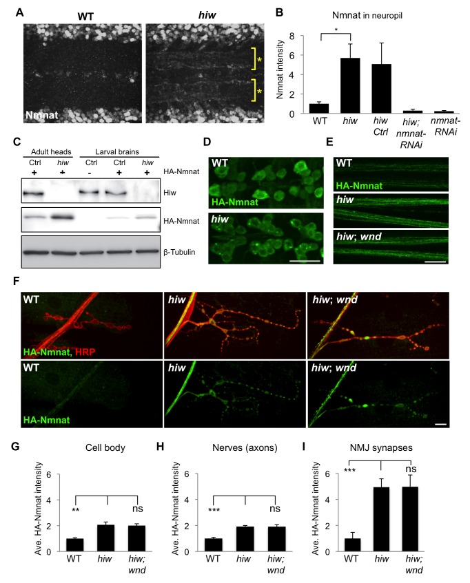
Figure 6. Hiw negatively regulates the levels of Nmnat protein in axons and synapses.
(A) Hiw regulates endogenous Nmnat protein in neuropil. In hiw ΔN mutants, Nmnat protein can be detected within the neuropil of the ventral nerve cord, denoted with asterisks. This area of the nerve cord is devoid of cell bodies and enriched in neurites and synapses. (B) Quantification (relative levels) of Nmnat staining in neuropil, for WT (w118), hiw mutant (hiw ΔN), hiw,Ctrl (hiw ΔN, BG380-Gal4), hiw, nmnat-RNAi (hiw ΔN, BG380-Gal4, UAS-nmnat-RNAi), and nmnat-RNAi (BG380-Gal4, UAS-nmnat-RNAi). See Materials and Methods. (C) Western blot with adult heads or larval brains to compare total protein levels of HA-Nmnat in wild-type (ctrl) and hiw mutant backgrounds. The UAS-HA::nmnat transgene is expressed in neurons with the BG380-Gal4 driver, and males are used for all experiments. (D–F) The UAS-HA-Nmnat transgene was expressed in motoneurons with the OK6-Gal4 driver, in wild-type (OK6-Gal4/UAS-HA::nmnat), hiw mutant (hiw ΔN;OK6-Gal4/UAS-HA::nmnat) and hiw; wnd double mutant (hiw ΔN ;OK6-Gal4/UAS-HA::nmnat;wnd1/wnd 2 ) backgrounds. HA-Nmnat protein is detected by immunostaining for HA. (D) Representative images of HA-Nmnat in motoneuron cell bodies, (E) segmental (peripheral) nerves, and (F) NMJ synapses, stained for anti-HA (green) and HRP (neuronal membrane, red). (G–I) Quantification of the average HA-Nmnat intensity for the above genotypes in (G) cell bodies, (H) segmental nerves, and (I) NMJ synapses at muscle 4. See Materials and Methods for details about quantification methods. In hiw mutants, Nmnat intensity is increased, particularly at NMJ synapses. Loss of wnd, in hiw;wnd double mutants, has no effect upon this increase. Scale bars = 12.5 µm, error bars represent standard error; *p<0.05; ***p<0.001; ns, not significant, p>0.05 in t-test.

