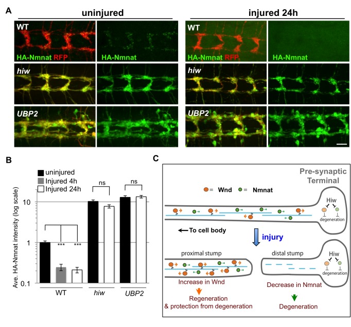
Figure 8. Hiw promotes Nmnat protein turnover in the injured distal axons and synapses.
(A) Distal axons and synapses of ppk-Gal4,UAS-mCD8::RFP labeled sensory neurons, located in the ventral nerve cord, either before or 24 h after injury. Transgenic HA::nmnat is expressed in a wild-type (WT) genetic background (UAS-HA::nmnat/+; ppk-Gal4,UAS-mCD8::RFP/+), hiw ΔN mutant (hiw ΔN ; UAS-HA::nmnat/+; ppk-Gal4,UAS-mCD8::RFP/+) or co-expressed with UBP2 (UAS-HA::nmnat/UAS-UBP2; ppk-Gal4,UAS-mCD8::RFP/+). Consistent with observations in motoneurons (Figure 6), mutation of hiw or co-expression of UBP2 causes a dramatic elevation in HA-Nmnat protein, and this does not disappear after injury, in dramatic contrast to the disappearance of HA-Nmnat in wild-type (WT) animals. Of note, the nerve terminals of the ppk-Gal4 labeled axons appear to be overgrown in hiw and UBP2 expressing mutants, similar to previous descriptions in other neuron types [8],[41]. Hence the quantification shown in (B) is normalized to the size of the nerve terminals (labeled by mCD8-RFP). Due to the expression of HA-nmnat, no axons are degenerating in any of the above genotypes. (B) Quantification of the average HA-Nmnat intensity in ppk-Gal4 expressing sensory neuron axon terminals, within the most posterior four segments of the ventral nerve cord. Error bars represent standard error. ns, not significant, p>0.05. (C) Model: Hiw promotes multiple independent responses to injury, through independent pathways. Hiw promotes degeneration in the distal stump by down-regulating Nmnat, and concurrently regulates regeneration (and protection from degeneration [15],[30]) in the proximal stump by regulating Wnd. Since Hiw may localize and function in distal axons, injury may relieve the inhibition of Wnd by Hiw in the proximal stump. Scale bars = 12.5 µm, error bars represent standard error; ***p<0.001; ns, not significant, p>0.05 in t-test.

