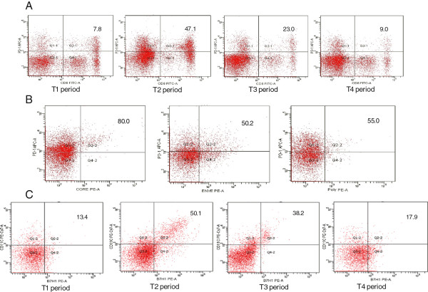
Figure 4.
(A) Representative dot plots of PD1 and CD8 double staining for patients in T1,T2,T3 and T4 sequence. Fresh heparinized peripheral blood samples were lysed with FACS lysing solution and then incubated with antibodies against PD1-APC and CD8-FITC for 20 min at 4°C. The numbers in the upper right quadrants indicate the frequency of PD1 in total circulating CD8+ cells. (B) Representative dot plots of PD1 and pentamer double staining for patients in T2 period. Fresh heparinized peripheral blood samples were lysed with FACS lysing solution and then incubated with antibodies against pentamer-PE and PD1-APC for 20 min at 4°C. The numbers in the upper right quadrants indicate the frequency of PD1-APC in circulating core18-27, enve335-343 and pol575-583 cells. (C) Representative dot plots of PD-L1 and CD11c double staining for patients in T1, T2, T3 and T4 sequence. Fresh heparinized peripheral blood samples were lysed with FACS lysing solution to remove RBCs and then incubated with antibodies against PD-L1-PE and CD11c-PEcy7 for 20 min at 4°C. The numbers in the upper right quadrants indicate the frequency of PD-L1 and CD11c dual positive cells.

