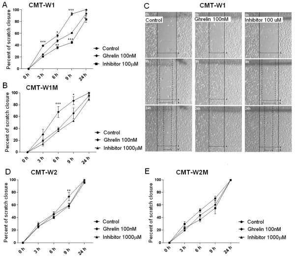
Figure 12.
Wound healing assay of canine mammary carcinoma cells after ghrelin treatment. Quantification of migration of CMT-W1 (A), CMT-W1M (B) CMT-W2 (D), CMT-W2M (E) cell lines represented as percentages of scratch closure and calculated as follows: % of scratch closure = a-b/a, where (a) is a distance between edges of the wound, and (b) is the distance which remained cell-free during cell migration to close the scratch. Ghrelin treatment at the concentration of 100nM increased cells motility after 9 and 24 hrs in the CMT-W1, CMT-W1M and CMT-W2 cell lines. Administration of ghrelin receptor inhibitor (100μM) decreased the motility of the CMT-W1 cells, whereas had no effect on motility of the cells of remaining cell lines. Photographs of cells invading the scratch were taken at the starting point and after 3, 6, 9, and 24 hrs using phase contrast microscopy (IX 70 Olympus Optical Co., Germany). (C) Representative pictures of scratch closure in control conditions, after ghrelin treatment (100nM) and GHS-R-specific peptide inhibitor [D-Lys3]-GHRP6 (100 μM) treatment in CMT-W1 cell line obtained using Olympus BX60 microscope (x4). Pictures have been analyzed using a computer-assisted image analyzer (Olympus Microimage™ Image Analysis, software version 5.0 for Windows, USA). The experiment has been conducted three times. Two-way ANOVA and Bonferroni post-hoc tests were applied. p < 0.05 was marked as *, p < 0.01 was marked as ** and p < 0.001 was marked as ***.

