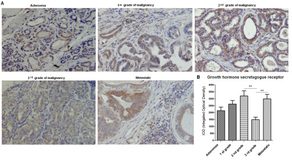
Figure 3.
Growth hormone secretagogue receptor expression in canine mammary tumors. (A) Light micrographs of canine mammary adenomas, adenocarcinomas of the 1st, the 2nd, the 3rd grade of malignancy and tumors that gave local/distal metastases (total n = 50) obtained with Olympus BX60 microscope (at the magnification of x20). The growth hormone secretagogue receptor (GHS-R) antigen is represented by brown colored precipitate in cytoplasm. (B) The graph of integrated optical density (IOD) of growth hormone secretagogue receptor (GHS-R)-positive cells in canine mammary tumors. The colorimetric intensity of the IHC-stained antigen spots was evaluated by a computer-assisted image analyzer (Olympus Microimage™ Image Analysis, software version 5.0 for windows, USA). Ten to 20 pictures in each slide were analyzed. The result are presented as a mean (±SEM) from 10 tumors in each group. The statistical analysis was performed using Prism version 5.00 software (GraphPad Software, California, USA). The one-way ANOVA and Tukey HSD post-hoc were applied to analyze the results. p- < 0.01 was regarded as highly significant and marked as **.

