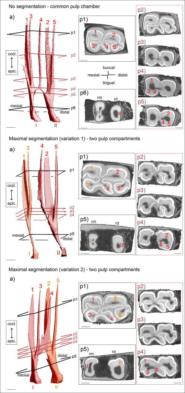
Figure 7.
Morphology of pulp horns and root canals in mandibular cheek teeth. Arabic numerals (1 to 7): Pulp horns. Roman numerals (I to III): Root canals. Colours indicate separate pulp compartments. (a) 3D images of the pulp cavity. Inserted planes p1 to p6 render the position of selected 2D μCT images, coloured planes indicate locations of connections. Sections (p1 - p6) demonstrate cross-sectional shape and size of pulp horns and root canals. Dark grey: Pulp tissue; light grey: Dentine and cementum; white: Enamel; rm = mesial root, rd = distal root. Scale bar: 5 mm. No segmentation (Triadan 409, 15 years): A narrow common pulp chamber connects all five pulp horns. Two distinct root canals are developed. Maximal segmentation (1) (Triadan 407, 7 years): Two pulp compartments are present, with pulp horn 4 included in the distal pulp compartment. Root canal I solely contributes to pulp horns 1 and 3. Root canal II solely contributes to pulp horns 2, 4 and 5. Maximal segmentation (2) (Triadan 407, 5 years): Two pulp compartments are displayed, with pulp horn 4 connected to the mesial pulp compartment. Root canal I solely contributes to pulp horns 1, 3 and 4. Root canal II solely contributes to pulp horns 2 and 5.

