Figure 1.
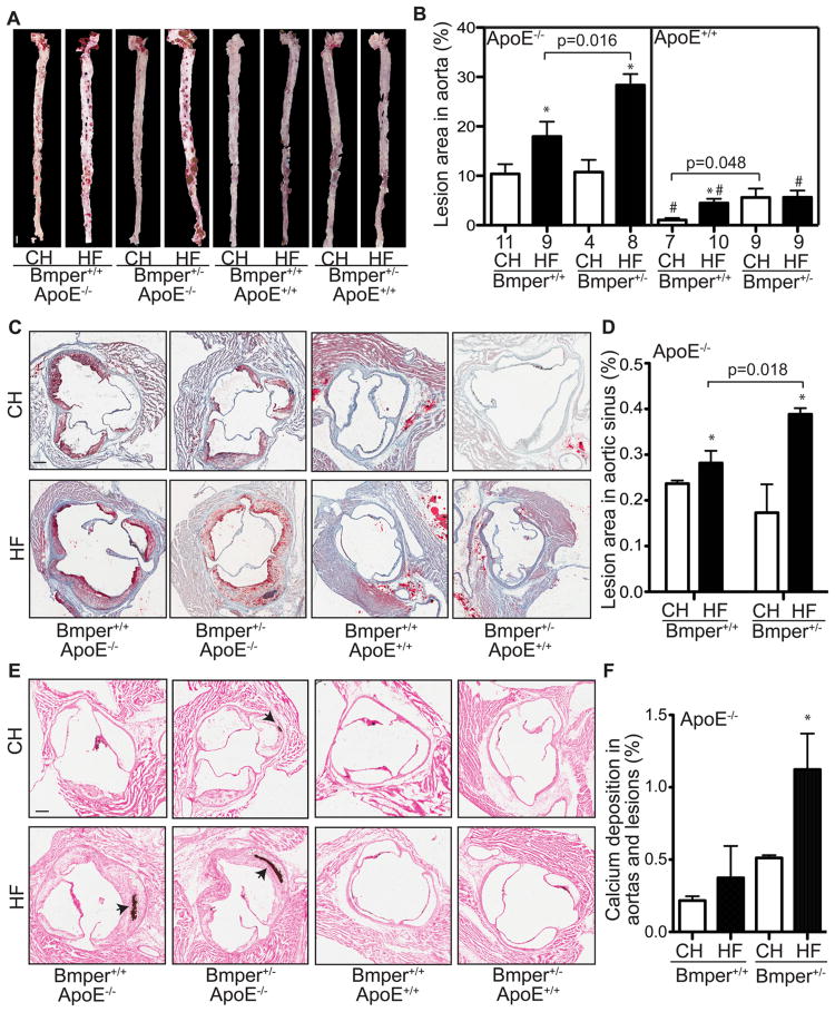
Bmper haploinsufficiency leads to aggravated atherosclerotic plaque formation and lesion calcification in ApoE−/− mice. Mice were fed a high fat diet (HF) or standard chow (CH) for twenty weeks. The aorta and heart were dissected out and stained with Oil Red O. A, Representative images of the Oil Red O staining of aortas. Scale bar, 1.5 mm. B. The lesions on the surface of each aorta were quantified as a percentage of the total area of the aorta. *, P<0.05, compared to mice with the same genotype but fed the control diet. #, P<0.05, compared to mice fed with the same diet but with the ApoE−/− genotype. The numbers below each column are the number of mice used in the experiments. C, Representative images of Oil Red O stained sections of aortic sinus regions from mice of the designated genotype and food groups. Scale bar, 0.2 mm. D, The lesions in the aortic sinus region were quantified as the percentage of the total lumenal area of aortas. *, P<0.05, compared to mice with the same genotype but fed the control diet, n≥4. There were no detectable lesions formed in the aortic sinus regions of ApoE+/+ mice. E, Representative images of calcification in the aortas and lesions as determined by Von Kossa staining (purple; indicated by black arrows). Scale bar, 0.2 mm. F, The area containing calcification deposition was measured as a percentage of the total lumenal cross-sectional area of aortas. *, P<0.05, compared to mice fed the same diet but with the ApoE−/− genotype, n≥5. No detectable calcification detected in aortas of ApoE+/+ mice.

