
Figure 1. Structures of the five norovirus complexes that are formed by full-length or truncated norovirus VP1.
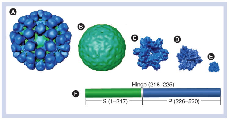
(A) Virus-like particle (180-mer: ~37 nm). (B) S particle (180-mer: ~27 nm). (C) P particle (24-mer: ~20 nm). (D)Small P particle (12-mer: ~14 nm). (E) P dimer (~6 nm). (F) Linear structure of norovirus VP1 with indications of the S (green) and the P (blue) domains that are linked by a short hinge. Numbers are based on Norwalk virus VP1.
P: Protruding; S: Shell.
Adapted with permission from [28].