
Figure 2. Formation and structural properties of norovirus P particles.
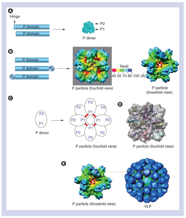
(A) The expression of the P domain with or without the hinge (left) forms a P dimer (right). (B) While the expression of the P domain (left) forms P particles (right), an end-linked C stabilizes the P particle formation. (C) P particle formation by P dimer. Red lines indicate the inter-P dimer disulfide bonds. (D)Fitting of the crystal structure of the norovirus (NoV) P domain into the density map of the P particle. P1 and P2 subdomains are indicated. (E) The NoV P particle shares similar surface antigenic structures to NoV. The VLP (right) is constituted by 180 VP1s with the P dimers on its outermost surfaces, while the P particle (left) is composed of 12 P dimers with similar orientation to those of the VLP. The two rectangles (red dashed lines) indicate the P2 region of a P dimer, representing the surface antigenic structures of both particles.
C: Cysteine-containing tag; P: Protruding; VLP: Virus-like particle.