
Figure 4. Structure of the protruding particle–VP8* chimera reconstructed by electron cryomicroscopy.
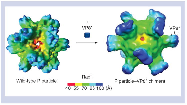
(A) The wild-type P particle before the VP8* antigen is inserted. (B) The P particle–VP8* chimera with the VP8* antigen on its outermost surface. Compared with the wild-type P particle, the chimera shows extended protrusions with nicks in the middle, suggesting the boundary between the P2 subdomain and the inserted VP8* antigen. The radii of the particles are indicated by the color schemes.
P: Protruding.
Adapted with permission from [16].