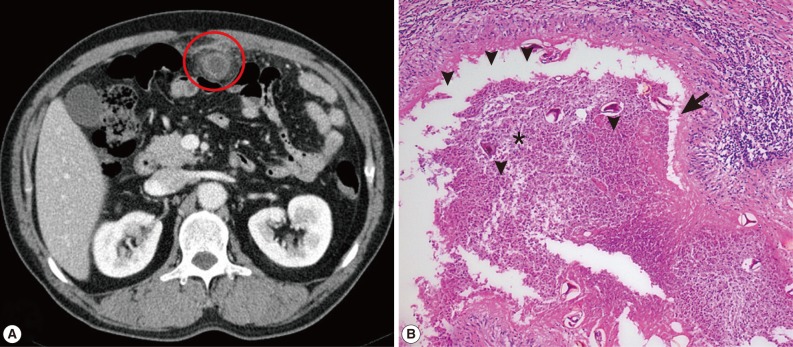
Fig. 1.
CT and microscopic findings of omental paragonimiasis. (A) Abdominal CT showed ring-shaped mass lesion with focal irregular enhancement in the omentum (red circle). (B) Microscopic pathologic examinations revealed the eggs of P. westermani (arrowheads) scattered with acute suppurative inflammations (star) and foreign body-type giant cells (arrow). H&E stain, ×100.

