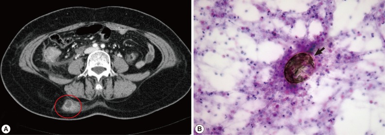
Fig. 2.
CT and microscopic findings of subcutaneous paragonimiasis. (A) CT scan shows localized abscess in the right back (red circle). (B) Fine needle aspiration cytology showed eggs of P. westermani (arrow) with eosinophil-dominant inflammatory cells. The eggs of P. westermani were yellowish-brown, ovoid or elongate, with a thick shell, and often asymmetrical with one end slightly flattened. H&E stain, ×400.

