Figure 6.
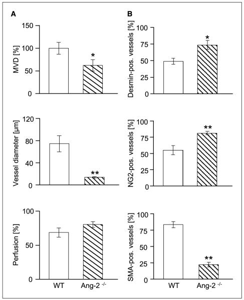
Effect of Ang-2 deficiency on MVD, vessel diameter and perfusion (A), and mural cell recruitment and maturation (B) in MT-ret melanomas. MT-ret melanoma cells were s.c. injected in WT and Ang-2–deficient mice and harvested when the first tumors had grown to 2 cm3. MVDs and vessel diameters were quantitated in CD31-stained tissue sections. Perfusion was assessed on the basis of FITC-lectin perfusion labeling. For mural cell coverage analysis, tumor sections were double-stained for the endothelial cell marker CD31 and for the mural cell markers desmin (top), NG2 (middle), and α-SMA (bottom). Vessel coverage was calculated as the percentage of desmin-, NG2-, and α-SMA–positive vessels compared with the number of CD31-positive vessels. As in LLC tumors, microvessels in MT-ret melanomas had higher desmin and NG2 coverage and lower αSMA coverage in tumors grown in Ang-2–deficient mice.

