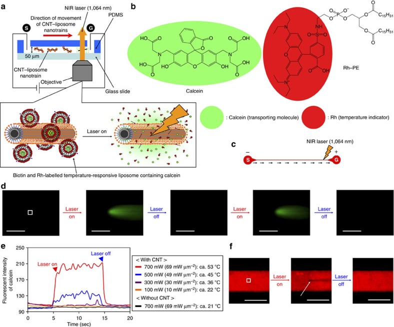
Figure 6. Molecular-transport system by CNT–liposome nanotrains.
(a) Concept of molecular transportation by CNT–liposome nanotrains. (b) Chemical structure of calcein and Rh–PE. (c) Design drawing of the microdevice based on a straight microchannel for molecular transportation. Black arrows show the direction of CNT–liposome nanotrain movement. S, start; G, goal. (d) Sequential calcein release by laser-induced-CNT–liposome nanotrains. The white dot square shows the location at which the fluorescence intensity of released calcein molecules was analysed. Magnification: X20. Applying voltage=100 V. Scale bars, 100 μm. (e) Fluorescence intensities of the released calcein molecules at various laser powers (100–700 mW (10–69 mW μm−2)). (f) A direct observation of ultrafast temperature change in the microchannel. The white dot square indicates the location at which the temperature was analysed in Fig. 4e. The white arrow indicates the fluorescence quenching of Rh by the photothermal property of CNTs. Magnification: X20. Laser power: 700 mW (69 mW μm−2). Applying voltage=100 V. Scale bars, 100 μm.

