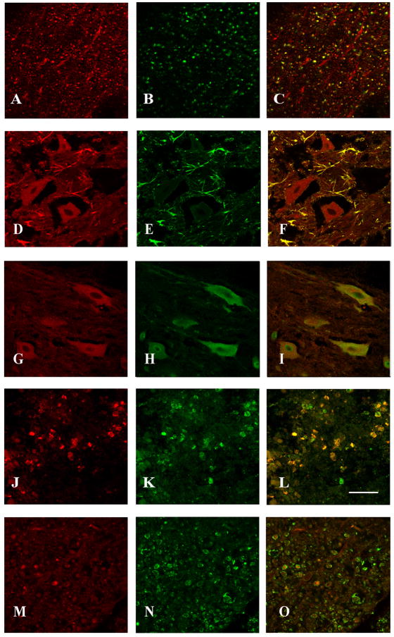
Figure 6.
Cell phenotype expressing ephexin in the injured spinal cord. Co-localization of ephexin in axons of the ventral white matter and in cells located in the gray matter such as astrocytes, neurons (close to the lesion epicenter) and macrophages (within the lesion cavity) was determined with double labeling studies and confocal microscopy (n=3). The first panel shows ephexin expression (red color; A, D, G, J), the second panel illustrates the different cellular/structural markers used (green color; NF-H (B), GFAP (E), NeuN (H), ED1 (K) and MAG (N)), and the third panel represents the merge (C, F, I, L). Ephexin (M) was not observed in myelin (N) structure of oligodendrocytes (O). Scale bar = 50μm

