
Figure 1.
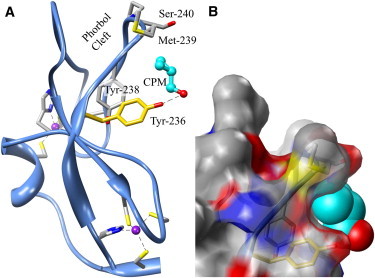
The overall structure of the PKCδ C1B subdomain complexed with CPM. (A) CPM (cyan) binds to the protein in a pocket created by Tyr-236, Asn-237, Tyr-238, Met-239, and Ser-240, and its hydroxyl group is hydrogen-bonded (dashed line) to Tyr-236 (2.8 Å). The site includes both Tyr-236 (gold), photolabeled by azialcohols, and part of the phorbol-binding site (Met-239, Ser-240) discovered by Hurley's group (31). Dashed lines are the coordination bonds to zinc (purple). (B) Surface representation in the same orientation as in A. Tyr-236, Tyr-238, Met-239, and Ser-240 are visible through the translucent surface. Nitrogen and oxygen atoms are blue and red, respectively. Molecular graphics images were produced using the UCSF Chimera package (59) (see Acknowledgments).