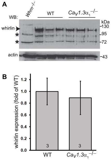
Fig 2. Whirlin expression level is not affected in the Cav1.3α1 null retina.
(A) Western blotting analysis shows no obvious difference in the whirlin expression level between the wild-type (WT) and Cav1.3α1 null (Cav1.3α1−/−) retinas. The arrow indicates the whirlin-specific band, and the asterisks indicate non-specific signals. The whirlin knockout retina (Whrn−/−) was used as a negative control. Actin signals on the same blot were shown in the bottom. The samples in different lanes are from different mice. (B) Quantitative analysis of whirlin signals on the western blot. The non-specific band at about 72 kDa on the whirlin blot was used as a sample loading control for each lane. The mean of the wild-type whirlin signals normalized by the sample loading control was defined as 1. The numbers at the bottom of each bar are numbers of animals examined. Error bars, the standard error of the mean.

