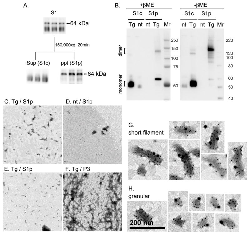
Fig. 4. Characterization of TBS-extractable 64 kDa tau.
A. TBS-extractable (S1) fractions were separated by further centrifugation to 150,000 × g supernatant (S1c) and precipitate (S1p) fractions, and 64 kDa tau was recovered from the S1p fraction. B. Reducing and non-reducing SDS-PAGE and Western blotting of S1c and S1p fractions. Reducing (+βME) and non-reducing (-βME) samples were subjected to Western blotting with the Tau46 antibody. Heat-stable S1c and S1p fractions from rTg4510 (tg) and non-tg (nt) 6 month-old mice were separated by SDS-PAGE on 4–12% Bis-Tris gels using MOPS SDS running buffer. C–H. Electron microscopy analysis with or without immunogold labeling. S1p fractions from rTg4510 (C) and non-tg (D) mice were immunogold labeled with anti-E1 (4 nm gold) and anti-Tau46 (10 nm gold). Conventional transmission electron showed short filaments in S1p fraction (E) and longer filaments in P3 fraction (F). In S1p fractions from rTg4510 mice, E1 and Tau46 antibodies labeled filamentous (G) and granular (H) structures. Scale bar is 200 nm.

