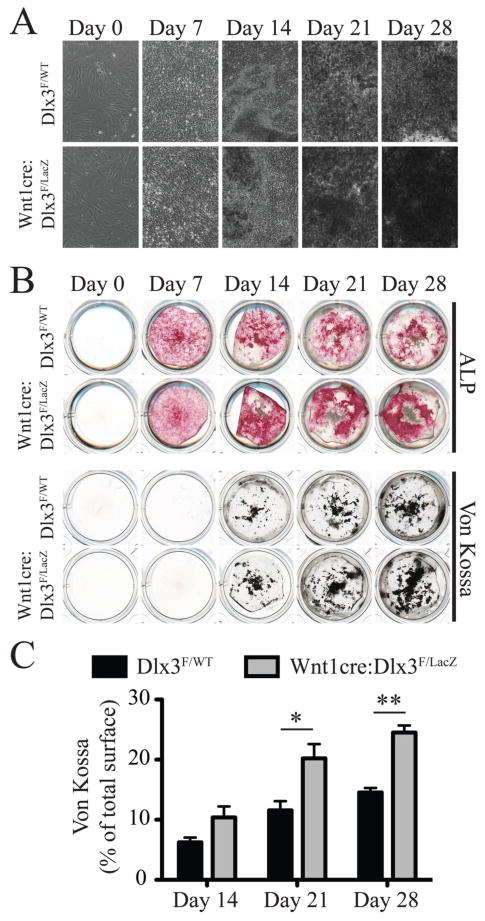
Figure 4. Ex vivo differentiation of osteoblasts isolated from neonatal frontal bones.
A) Calvaria cell cultures isolated from frontal bones from WT and cKO animals. Images were acquired 0, 7, 14, 21 and 28 days after induction of osteoblast differentiation using osteogenic medium. Images at days 14, 21 and 28 were taken in areas showing the highest nodule density. B) Alkaline phosphatase (ALP, upper panel) and Von Kossa (upper panel) stainings of calvaria cell cultures described in A. C) Quantification of the relative surface of Von Kossa staining (percentage of total well surface) for WT and cKO cell cultures at 14, 21 and 28 days after induction of differentiation.

