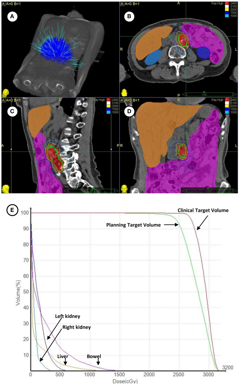
Figure 1.
(A) Depicted are the 140 treatment beams by the Cyberknife radiosurgery 6 MV accelerator for treatment during the targeting of left-sided para-aortic lymph nodes. (B–D) Depicted are axial, coronal, and sagittal projections of radiosurgical treatment. The 18F-FDG PET/CT-derived clinical target volume (red shaded volume) and 3 mm expanded planning tumor volume (green shaded volume) are contoured. The 24 Gy prescription isodose is highlighted in red and a 10 Gy isodose is outlined in light blue. Contouring of the bowel (magenta), liver (orange), right kidney (light blue), and left kidney (dark blue) is shown. (E) Plotted are corresponding radiation dose-volume histograms for clinical targets and organs at-risk.

