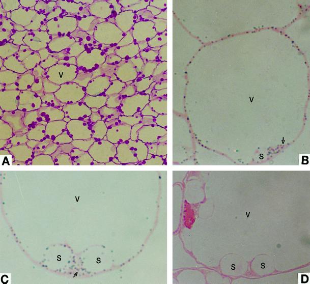
Figure 1.
Light micrographs showing immunolocalization of AGP in developing pericarp of tomato fruit. A, Carbohydrate-specific staining. Starch grains stained red. B and C, Anti-tomato fruit AGP serum detected a signal (gray dots) in both plastids and cytoplasm (arrows). D, The preimmune control serum detected no signal. S, Starch granule; and V, vacuole. A, ×351; B, ×1560; and C and D, ×1950.

