Figure 2.
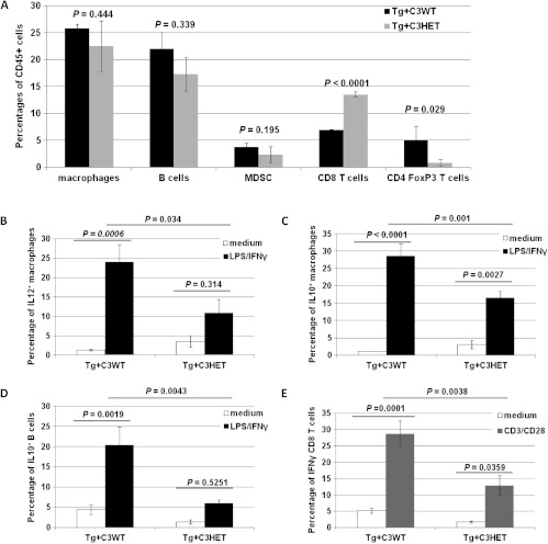
Functional impairment of tumor-immune infiltrate in Tg+C3HET mice. (A) Ovarian tumors from 16-week-old Tg+C3WT mice (black bars; n = 4) and Tg+C3HET mice (gray bars; n = 4) were dissociated and stained for CD45, CD11b, B220, F480, GR1, CD3, CD8, CD4, CD25, and FoxP3. CD45+ leukocytes were gated and macrophages were identified as CD11b+F480+, B cells as CD11b-B220+ cells, MDSCs as CD11b+GR1+, CD8+ T cells as CD3+CD8+ cells, and regulatory T cells as CD4+CD25+FoxP3+. Bars represent percentages of CD45+ cells ± standard error. (B-E) Ovarian tumors were collected from 16-week-old Tg+C3WT (n = 4) and Tg+C3HET (n = 4) mice, mechanically dissociated to a single-cell suspension and incubated 4 hours in medium only (white bars), or activated for 4 hours with LPS and IFN-γ (black bars), or with anti-CD3 and anti-CD28 mAbs (gray bars). Cells were then stained for CD45, F480, and CD11b (macrophages) (B, C), CD45 and B220 (B cells) (D), or CD45, CD3, and CD8 (T cells) (E), washed and permeabilized for intracellular staining for IL-12 (B), IL-10 (C, D), or IFN-γ (E). Bars represent mean percentages ± standard errors.

