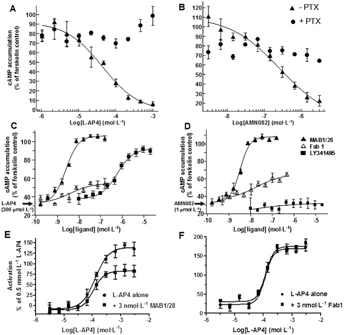
Figure 9.

Functional characterization of MAB1/28 and Fab1 fragments in cAMP and [35S]-GTPγ binding assays. Concentration–response curve of L-AP4 (A) and AMN082 (B) in the absence and presence of PTX in the CHO mGlu7 expressing stable cell line 83. Cells were stimulated with 3 µmol·L−1 forskolin, and various concentrations of L-AP4 or AMN082. Concentration–dependent inhibition of 300 µmol·L−1 L-AP4 (C) and 1 µmol·L−1 AMN082 (D) by MAB1/28, Fab1 fragments or LY341495 in the CHO line 83 cells. Cells were stimulated with 3 µmol·L−1 forskolin, an EC80 of L-AP4 or AMN082 and various concentrations of MAB1/28, Fab1 or LY341495. The cellular content of cAMP was measured and expressed as % cAMP content compared to cells treated with 3 µmol·L−1 forskolin only. Concentration–response curves for L-AP4-induced [35S]-GTPγ binding in the membranes from CHO mGlu7 cell line 83, in the absence and in the presence of 3 nmol·L−1 MAB1/28 (E) and in the absence and in the presence of 3 nmol·L−1 Fab1 fragments (F). All measurements were performed in triplicate and values represent mean ± SEM.
