
Figure 6.
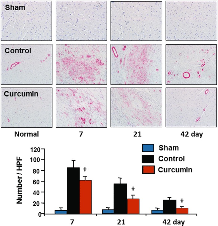
Differentiation of fibroblasts to myofibroblasts. No α-SMA expressing myofibroblasts were detected in the non-ischaemic zone in the control group or in either of the sham groups. Ischaemia/reperfusion caused a significant increase in the number of α-SMA expressing myofibroblasts accumulated during reperfusion, which subsequently declined to day 42 in the control group. However, this increase in accumulation of α-SMA expressing myofibroblasts was significantly reduced by curcumin relative to each time point during reperfusion in the control group. n= 8, values are mean ± SEM. *P < 0.05 versus sham; †P < 0.05 versus control.