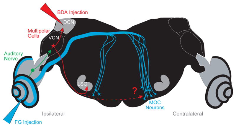
Figure 1.

Neural pathway of the medial olivocochlear (MOC) neurons projecting to the left cochlea (Ipsilateral side). Also shown are the stages of the reflex pathway leading to the MOC neurons: the auditory nerve fibers that project to the ventral cochlear nucleus (VCN), and the VCN multipolar cells that presumably project to MOC neurons. To test the hypothesis that multipolar cells project to MOC neurons, we labeled them via their collaterals using biotinylated dextran amine (BDA) injected into the dorsal cochlear nucleus (DCN). Multipolar cell axons were followed into the superior olivary complex (SOC), where many form branches to the lateral superior olive (LSO) on the ipsilateral side as reported previously (Doucet and Ryugo, 2003). Results of the present study show that they also form branches to the ventral nucleus of the trapezoid body (VNTB) on the contralateral side, the location of most MOC neurons in the mouse. In some experiments, a second injection of Fluorogold (FG) was made into the cochlea to retrogradely label MOC neurons.
