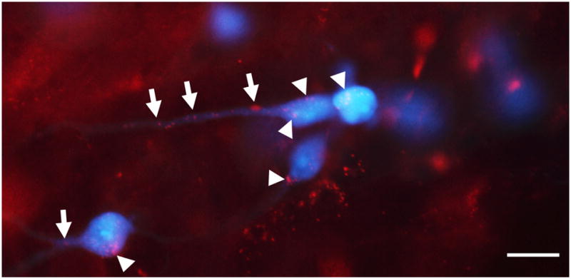
Figure 7.

Light micrograph of the contralateral VNTB showing boutons of presumed multipolar cells (red) that contact MOC neurons (blue) on their somata (arrowheads) and dendrites (arrows). In this double-injected case, fluorescently tagged DA (red) was injected into the DCN and Fluorogold (blue) was injected in the cochlea. Scale bar = 15 μm.
