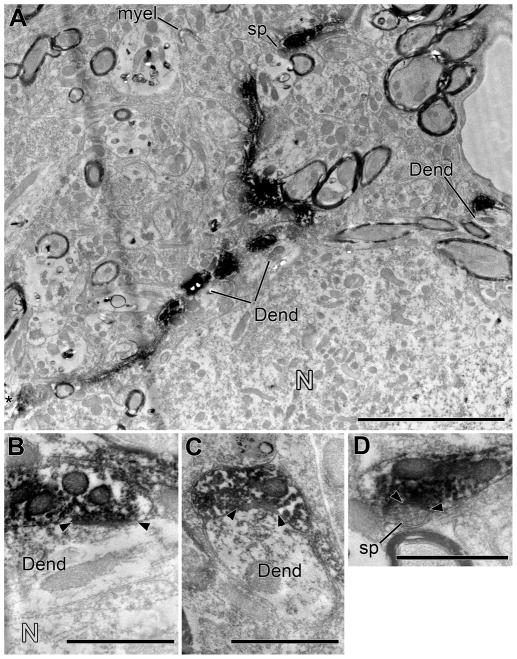
Figure 8.
Electron microscopy of a labeled branch from a different case than Figure 6. A: Low-magnification electron micrograph shows a labeled branch winding through the contralateral VNTB neuropil adjacent to a neuron (N). The neuron was not a postsynaptic target in our material, but nearby postsynaptic targets included two dendrites (Dend, one of them indicated in two segments in the center and confirmed to be contiguous in serial sections) and a spine (sp). In the neuropil, an axon of unknown origin forms a large ending as it loses its myelin (myel). Scale bar = 5 μm. B–D: Higher-magnifications of the synapses on dendrites (Dend) and a dendritic spine (sp). Panel B reveals a dendrite intervening between the labeled branch and the neuron. Scale bars = 1 μm.

