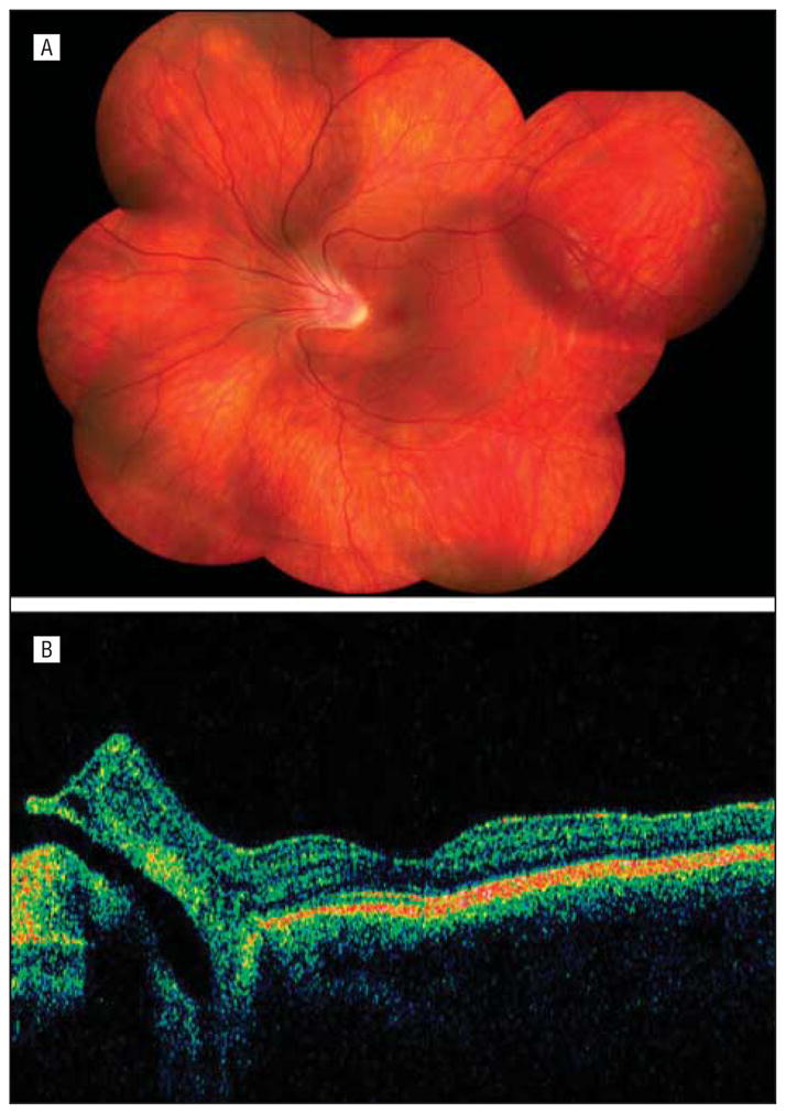
Figure 2.

Individual 8. A, Composite fundus photograph of the left eye shows an optically empty vitreous with preretinal vitreous condensation in the midperiphery, avascular vitreous sheets in the far periphery, and nasal dragging of the arcades and fovea. B, An optical coherence tomogram of the left eye shows the optic nerve inverted nasally.
