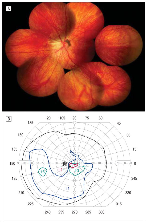
Figure 7.
Individual 11. A, Composite fundus photograph of the left eye shows an optically empty vitreous except for a subtle midperipheral avascular vitreous membrane, mild peripapillary pigmentation, straightening of the vascular arcades, and subtle inferior chorioretinal atrophy. B, Goldmann visual field test of the left eye shows early superior scotomatous changes that correspond to the inferior chorioretinal atrophy seen on fundus photography.

