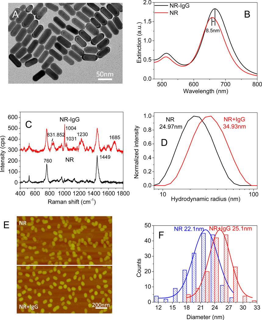
Figure 2.
(A) TEM image of AuNRs. (B) UV-vis extinction spectra confirming AuNR and SH-PEG-IgG conjugation. The UV-vis extinction spectrum of 56×22 nm AuNRs shows a longitudinal LSPR wavelength of 658 nm (AuNRs, red). After incubation with SH-PEG-IgG, the λmax red shifts 8.5 nm (AuNR-IgG, black). (C) Hydrodynamic diameters obtained from DLS show that the average hydrodynamic diameter increased by 10nm following IgG conjugation. (D) Surface enhanced Raman spectra before and after the conjugation of antibody on gold nanorods reveal the Raman bands corresponding to IgG. (E-F) AFM images of AuNRs and AuNR+IgG conjugates, revealing the increase in the average diameter of the nanorods following the bioconjugation.

