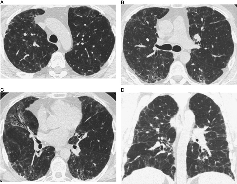
Figure 1.
High-resolution CT (HRCT) images of interstitial abnormalities of nontypical CT scan pattern show bilateral patchy areas of ground-glass opacity superimposed on fine reticulation, distributed diffusely in the craniocaudal and axial planes. A, B, and C, Transverse thin-section CT scans. D, Coronal scan.

