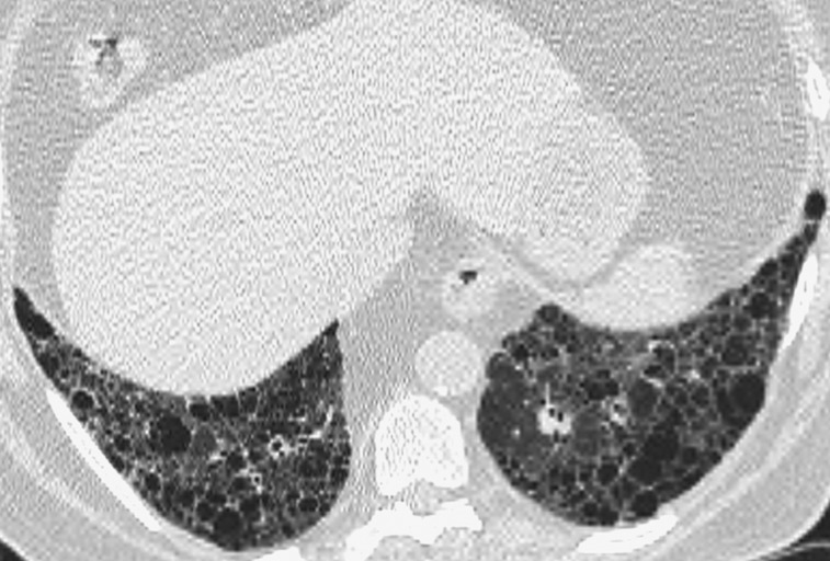
Figure 2.
Representative abnormal HRCT image of definite usual interstitial pneumonia pattern. Transverse thin-section CT scan of basal segments of lower lobes shows reticulation and honeycombing. See Figure 1 legend for expansion of the abbreviation.

