
Figure 3.
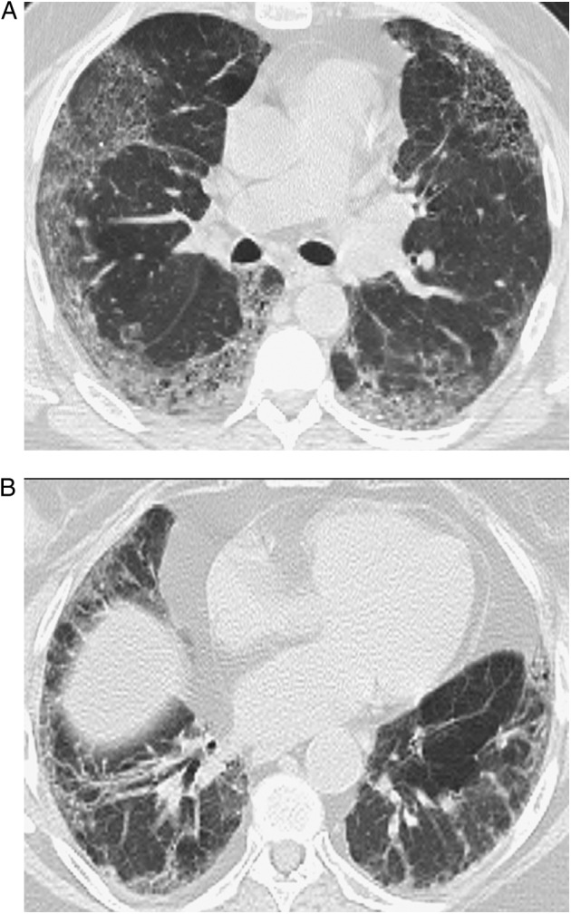
A, B, Representative abnormal HRCT images of probable nonspecific interstitial pneumonia pattern. Transverse thin-section CT scans of basal segments of lower lobes show peripheral predominant ground-glass opacity with mild reticulation. See Figure 1 legend for expansion of the abbreviation.