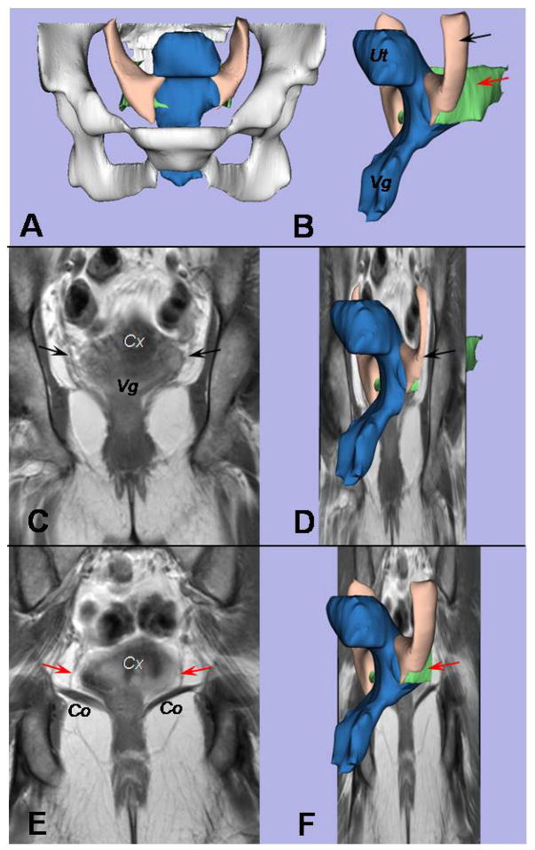
Figure 2.
A coronal view of the cardinal and uterosacral ligaments is provided. A, The bony pelvis can be seen in this front view of the 3D model. B, An oblique left-side view displays the uterus (Ut) and vagina (Vg) in blue, the uterosacral ligament in green (red arrow), and the cardinal ligament in cream (black arrow). C, A coronal scan is at the level of the cardinal ligament; the cervix (Cx) and vagina (Vg) are marked. D, This is where the structures in the 3D model shown in Figure 2B would be situated in the plane. E, The uterosacral ligament (red arrows) can be seen when the view is moved 10 mm dorsal from where the cardinal ligament is seen in Figure 2C. The cervix (Cx) and coccygeus muscle (Co) are shown. F, Again, the 3D model shown in Figure 2B is shown within the coronal plane.

